MAHI

Multimodal Animal Health Informatics

University of Wisconsin–Madison

Building AI for patients who can't tell us what's wrong.

Six research images side by side: a scatter of coloured points from a genome embedding, a pink and purple stained tissue tile, a three-dimensional reconstruction of a heart specimen on black, a retinal scan with vessels traced in green and the optic disc outlined in orange, a facial landmark graph drawn in cyan over a video frame of a dog, and an infrared night camera-trap frame of a deer.
From molecules to populations — genome embeddings, histopathology, gross specimens, clinical imaging, behaviour, and wildlife surveillance.

About

MAHI is a multidisciplinary research community at the intersection of computer science, clinical veterinary medicine, and health informatics.

Frontier clinical AI has been built almost entirely around human patients. MAHI works to close that gap — adapting modern methods to the species-specific biological constraints of animal patients, and bringing companion, production, and wildlife animal health problems to the AI research community in a form it can work with.

We synthesize multimodal data — genomic, imaging, clinical, and environmental health signals — into informatics frameworks supporting early disease detection, wildlife surveillance, and clinical decision support, with an emphasis on transparent, safety-first systems that improve outcomes for companion, wildlife, and production animals.

Focus areas

  • Clinical & companion animal

    Decision support, diagnostics, clinical records.

  • Production & dairy systems

    Herd-scale monitoring and early detection.

  • Wildlife & population health

    Surveillance under sparse, opportunistic sampling.

  • Trustworthy AI

    Evaluation, validation, and transparency for animal health.

The kind of work

Animal health data arrives in many forms — camera-trap frames, video, whole genomes, whole-slide images, gross specimens, and clinical imaging. The examples below reflect the data currently at hand, not the boundaries of what the group is interested in.

Six white-tailed deer photographed by camera traps, ordered by model score from 0.05 to 0.90. The first three are outlined in blue and labelled not low; the last three are outlined in orange and red and labelled flag, low. A blue-to-red colour bar marks the flag threshold at 0.50.
Wildlife surveillance Body-condition scoring on Snapshot Wisconsin camera-trap frames. Each usable frame gets a probability that the animal is in low condition, and anything past the threshold is flagged for a person to look at. It surfaces suspects for review; it does not diagnose.
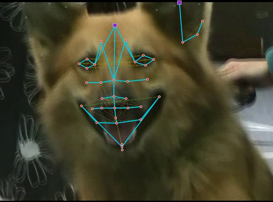
A video frame of a German shepherd's face with a facial landmark graph drawn over it: red nodes joined by bright cyan anatomical contours, fainter orange shared-muscle edges, and purple square super-nodes at the ears and pose reference.
Behaviour A facial landmark graph tracked frame by frame through video. Cyan edges follow anatomical contours, fainter edges encode shared musculature. Scoring is gated on pose and landmark uncertainty — frames it cannot read are abstained on rather than guessed.
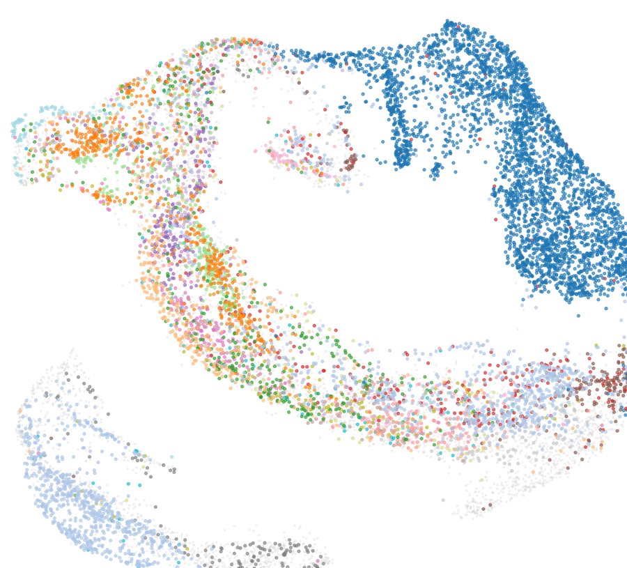
A UMAP scatter plot of canine genome embeddings. Thousands of small coloured points form arcs and lobes, with one large tightly clustered blue group at the upper right.
Genomics A genome foundation model trained on 14,478 canine genomes across 386 breeds. Each point is one animal, coloured by breed — the model was never shown a breed label.
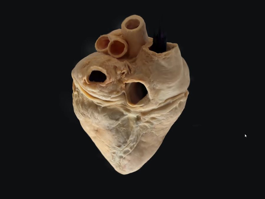
A three-dimensional reconstruction of a gross heart specimen rendered against a black background, with great vessels visible at the top.
Gross pathology Gaussian splatting of a gross specimen — photographed from every angle, then reconstructed as a 3D model that can be rotated, relit, and measured after the specimen itself is gone.
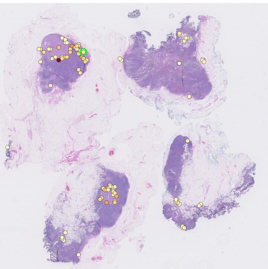
Four pale lavender tissue sections on a whole-slide image, scattered with small yellow and orange dots marking where a classifier placed attention, densest over the darker tumour regions.
Whole-slide imaging Where a slide-level classifier places its weight as it traverses a whole-slide image. Attention concentrates over the darker tumour regions rather than spreading evenly across the tissue.
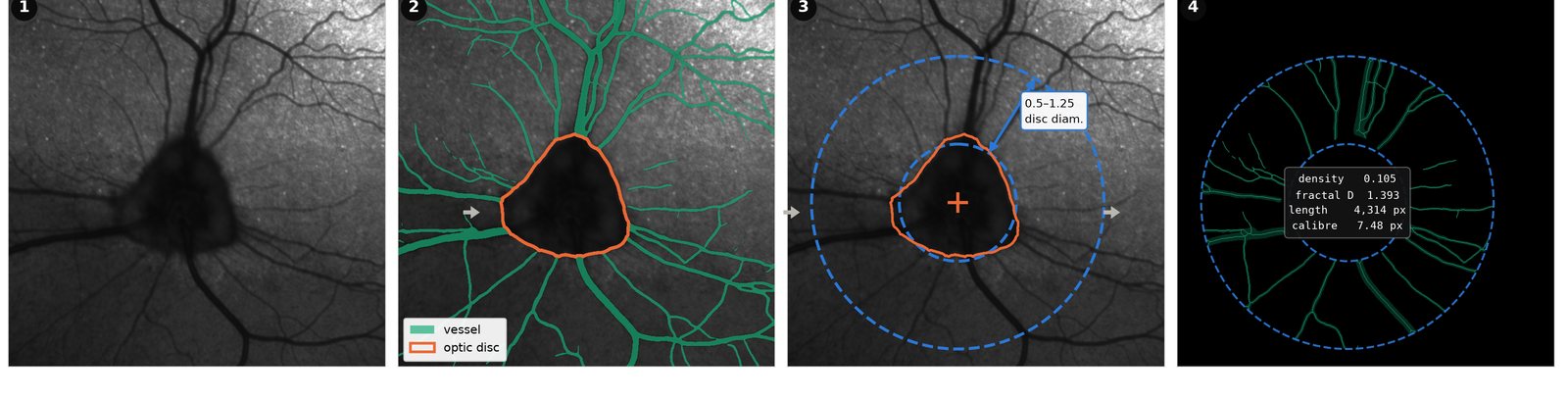
Four panels of a retinal imaging pipeline: an acquired infrared en-face frame, the same frame with vessels and the optic disc segmented, a region of interest anchored to the disc in disc-diameter units, and the extracted vascular skeleton with density, fractal dimension, length and calibre measurements.
Clinical imaging Retinal vascular structure measured in awake animals, where the eye moves between frames. Segment, anchor the region to the optic disc, measure every frame, then average.

Figures from research at the UW School of Veterinary Medicine.

Fall 2026

MAHI's first semester is focused on community building. We are convening researchers from veterinary medicine, animal and dairy sciences, wildlife health, computer science, and informatics through a kickoff networking lunch, a biweekly problem-and-methods lunch series, and a half-day ideation workshop.

If you work on animal health data, or on methods that might apply to it, we'd like to hear from you.

Who's involved

Participants come from across the UW–Madison campus and partner organizations, including:

  • Department of Surgical SciencesSchool of Veterinary Medicine
  • Department of Medical SciencesSchool of Veterinary Medicine

Get involved

Email
Brundage2@wisc.edu
Mailing list
Sign up form (opens in a new tab)